Focal Extender System

 
Focal Extender:  Combine The Best Focus From Multiple Focal Planes And Improve Your Image
FROM THIS SINGLE PLANE FOCUS...
TO THIS!
Page Contents
Sample Images:
Firebeetle Infrared Sensor
Histological
Other Sample Images:
Neurological

Information:
What Is It?
Advantages Over Confocal Microscopes
Why Is It Needed?
How Does It Work?
What Is Included In The Extended Focus System?
Frequently Asked Questions

Actions:
I'd Like More Information/Email Us
Go To Our Homepage


Sample Images
Below are some sample images generated from the Focal Extender application. 
These images were acquired using brightfield lighting on a compound microscope.

For each sample, there are 4 images:

The 'Typical Single Plane Image' shows what you would normally see through the microscope or when imaged.
The 'Focal Extender Image' shows what the image looks like after the Focal Extender application is used.
The 'Actual Focal Plane Movie' shows what is seen through the microscope as the focus is moved through the sample.
The 'Sampled Rotational 3D Movie' gives a sample of the 3D rotation that can be visualized after the images are collected.
 
Infrared Sensor From A Firebeetle
These images were acquired using fluorescence on a stereo microscope.
Typical Single Plane Image

Download Full Resolution Image

Focal Extender Image

Download Full Resolution Image

Actual Focal Plane Movie

Download High Resolution Movie

Sample Rotational 3D Movie

Download High Resolution Movie

 
Histological Samples
These images were acquired using brightfield on a compound microscope.
Typical Single Plane Image


Focal Extender Image


Actual Focal Plane Movie


Sample Rotational 3D Movie


What is it?
The Focal Extender application combines the 'best focused' portions of multiple image focal planes to generate a single 'in-focus' image, thereby increasing the depth of field of your microscope by up to 100 times.  Instead of only having a portion of the field of view focused, you can now have the entire field of view in focus at one time in a single image, vastly improving the visual quality and usefulness of your images.

If you are familiar with confocal microscope systems, basically this is a MUCH less expensive way of obtaining results similar to a confocal microscope. 


Advantages Over Confocal Microscopes
Price
For about 10%-30% of the cost of a confocal system, you can produce very similar results.

Works in BOTH BRIGHTFIELD and FLUORESCENCE

You can produce images with both brightfield and fluorescent samples, not just fluorescent as with a confocal system.

Easy To Use

Unlike confocal systems, which can be very difficult to work with, it is very easy to obtain and work with your images.

Less Finicky

You don't need to worry about getting the system set to very exacting settings to capture your images.  There aren't a lot of settings to get adjusted perfectly before you capture your images.  All you need to do is set your lighting properly and adjust the camera settings.

Can Be Used With Stereo Microscopes

We have had many users inquire about using the system with stereo microscopes in order to image larger samples at lower magnifications with a wider field of view than is possible with a compound microscope.  The Extended Focus system will work with some stereo microscopes, unlike confocal systems which use only research microscopes.

 
Why is it needed?
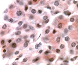
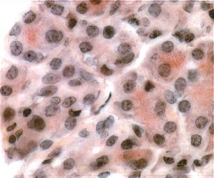
Very often when taking images (especially when imaging thicker samples) only some portions of the field of view are in focus for any particular focal plane, the rest of the image is out of focus.  Previously, researchers had to select which portion of the image would be in focus and suffer from the rest of the image being out of focus. 

Now researchers are able to obtain a single image where the entire field of view is in focus at one time allowing for visualization and publication that more clearly represents the actual sample! 

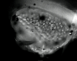
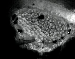
An additional benefit is that once the series of focal planes is acquired, a 3 dimensional model can be created allowing you to see your sample through any rotational angle desired, enabling you to see your sample as never before (see the sample images above for examples).


How does it work?
By motorizing the focus of the microscope, we acquire images at multiple focal planes that are equally and closely spaced.  For each pixel in the Extended Focus image, the software looks at all of the images in your various focal planes and takes the information from the focal plane where that pixel has the best focus.  This is done for each pixel in your image so that when finished, all of the 'best focus' pixels from your image planes are then combined creating an 'extended depth of field' image composite allowing you to see much more of your field of view in focus at one time than is normally possible.

 
What is included with the system?
There are several components which we supply that comprise the Extended Focus system:
1.)  A digital camera with very high optical resolving capabilities.
2.)  A motorized focus control.  If you have a built in focus control, we can possibly use it, depending on your microscope model.  Or we can add a motorized focus control to most microscopes if you do not have it built in.
3.)  Software to collect the images, generate the 3D visualizations and the Extended Focus image.
4.)  The adapters to fit the camera and motorized stage to your microscope.
5.)  Optionally, we can provide the computer system as well.

 
Frequently Asked Questions
Q:  What types of microscopes does it work on?

A:  Just about any type.  We support compound (research) microscopes from Leica, Nikon, Zeiss, and Olympus.  We also support stereo microscopes from Leica.

Q:  Can it bring into focus portions of the field of view that are not in focus in any of the image planes acquired?

A:  No.  We can only project the 'best focused' portions of the image planes.  In order to obtain focus in the resultant image, the object must be in focus in at least one of the focal planes.

Q:  Is it easy to use?

A:  Absolutely.  You only need to select the uppermost and lowermost focal planes and the number of images you want to take.  The rest of the procedure is automatic.

Q:  Does it work with brightfield or fluorescence images?

A:  Both.

Q:  Does it work in color or monochrome images?

A:  Both.


I'd Like More Information
Will It work with my images?
If you would like to know if the system would work with your images, please feel free to email us a sample image.  Please send the image in TIF or JPG format and limit the file size to 1 Megabyte or less.

Will It work with my microscope?

If you would like to know if the system would work with your microscope, please include information on:
1.)  The manufacturer and model of your microscope.
2.)  If you have a built in focus control, please let us know.
3.)  If you have a 3rd party focus control, please let us know the manufacturer and model.

Email us


Return to Home Page